Respiratory model
The Pulse Physiology engine is designed to model the ventilatory behavior of a patient Respiratory System. An electric analog lumped parameter model was used to do so.
Let’s analyse the modelisation of the human respiratory system, and pull out its main features.
First of all, in order to breath, we need air, found in our environment. Therefore, our respiration depends heavily on the conditions of the environment within which our body is located. Environment conditions are taken into account in the Ambient Environment section of the Respiratory Model.
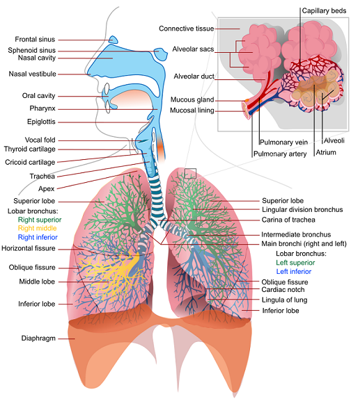
The air we breath is pumped in and out our body through a system of pipes and goes into what we call the respiratory tract. The latter is divided into upper and lower airways. The upper airway consists of the region above the cricoid cartillage, whereas the lower airways begin at the trachea and extend to the bronchi, bronchioles, and the alveoli.

The two openings of the airway (the nasal cavity and the mouth) meet at the pharynx. The pharynx has the ability to carry both food and air. At the bottom of it, the pathway divides into two sections: the esophagus (for food only) which leads to the stomach, and the trachea (for air). Once the air is in the trachea, it goes down to the carina, intersection that branches to form the right and left main bronchi. Then, the bronchi bifurcate into smaller bronchioles that continue branching for up to 23 generations, forming the tracheobronchial tree that terminates with the alveoli.
In the tracheobronchial tree, only a small portion of the branches participate in gas exchanges. We distinguish the conducting zone from the respiratory zone.
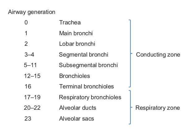
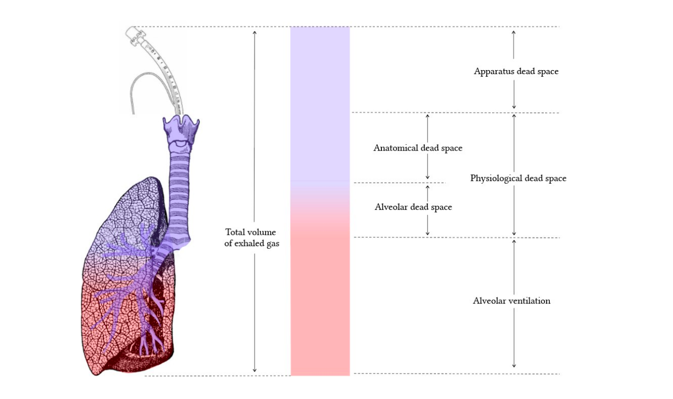
The first several generations of airways, where no gas exchange occurs, constitute the anatomic dead space and are referred to as the conducting zone. Their function is to further warm, moisten, and clean the inspired air and distribute it to the gas-exchanging zone of the lung.
The gas-exchanging zone of the lung corresponds to the respiratory zone, where alveoli are present. This section includes respiratory bronchioles, alveolar ducts, alveolar sacs, and alveoli.
Not all alveoli have the ability to perform gas exchanges. These alveoli are part of the alveolar dead space.
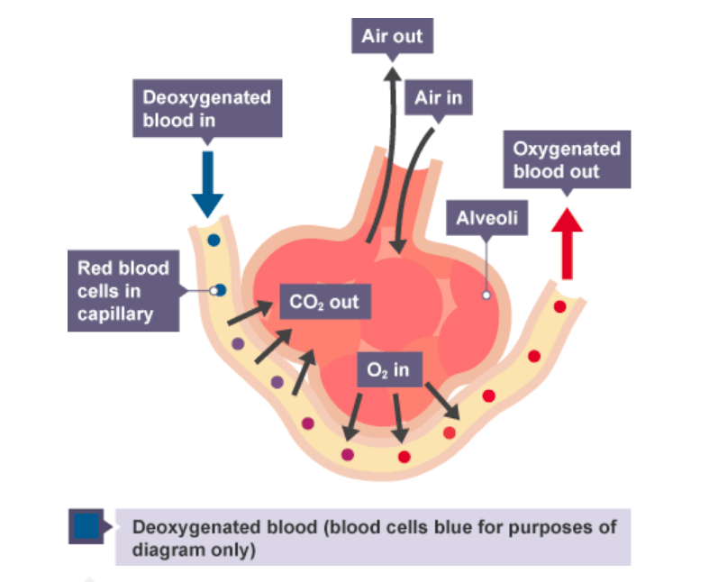
The respiratory zone is the site of oxygen and carbon dioxide exchange with the blood. The respiratory bronchioles and the alveolar ducts are responsible for 10% of the gas exchange. The alveoli are responsible for the other 90% that occurs between the airways and the Cardiovascular System. During gas exchange, oxygen moves from the lungs to the bloodstream, and carbon dioxide passes from the blood to the lungs. It happens in the lungs between the alveoli and tiny blood vessels called capillaries, located in the wall of the alveoli.
The alveoli have the specificity to inflate when a person inhales and deflate when a person exhales. Inhalation and exhalation are helped by the respiratory muscles, and in particular by their elasticity. In the model, these features are represented by capacitors (that represent compliance).
During the respiratory process, insults can happen and lead to gas leak. The gas leak can come either from the alveoli (alveoli leak) or from the thoracic wall (chest leak). It is the reason the model accounts for the pleural cavity through circuit elements that allow flow into the pleural space.
The respiratory system is modeled thanks to the following electric analogue circuit:
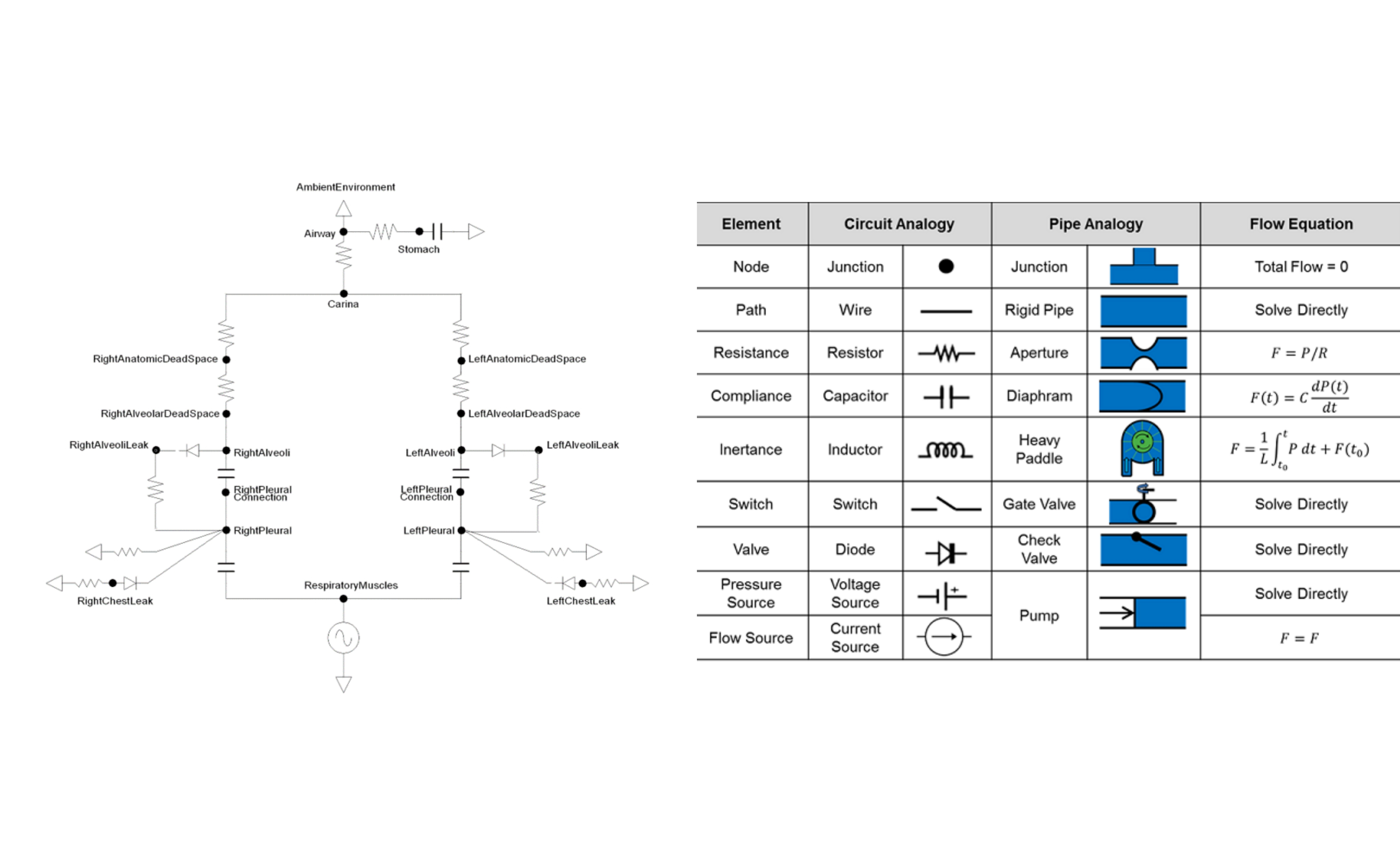
References
-
[CO2] Rebecca Lindsey Climate Change: Atmospheric Carbon Dioxide August 14, 2020
-
[C02_emissions] STEPHANIE OSMANSKI How Do Carbon Emissions Affect the Environment? MAR. 30 2020
-
[energy_in_buildings] Why are household energy efficiency measures important for tackling climate change? 5 June, 2018
-
[Ibat] Christophe Prudhomme 4fastsim-ibat OCTOBER 21, 2019
-
[IAQ] Indoor Air Quality
-
[IAQ_covid] Indoor Air and Coronavirus
-
[pulse] Pulse
-
[Respiratory_Methodology] Pulse respiratory
-
[low_airway] Cambridge University Press The Lower Airways 2019
-
[dead_space] Dead space and its components
-
[respiration] Respiration
-
[normal_pulse] Pulse & Heart Rate
-
[blood_pressure] Blood pressure
-
[med_ill] Medical illustration
-
[end_tidal_carbon_dioxide] End-Tidal CO2 May 2018
-
[lung_vol] Lung volumes
-
[skin_temp] Skin temperature
-
[rep_sys_list] Respiratory System List
-
[altitude_hr] Elevated Thinking: Altitude and the Heart Mar 30, 2012
-
[lung_ha] The lung at high altitude
-
[everest_ha] Robert Preidt Everest Study Finds High Altitude Affects BP Aug. 27, 2014
-
[thin_air] Living in thin air
-
[body_temp] Body temperature
-
[covid_mask] Mask
-
[ventilator] Jeffrey B. Webb ,Aaron Bray,Philip K. Asare,Rachel B. Clipp,Yatin B. Mehta,Sudheer Penupolu,Aalpen A. Patel,S. Mark Poler Computational simulation to assess patient safety of uncompensated COVID-19 two-patient ventilator sharing using the Pulse Physiology Engine November 25, 2020